Brain Scans Link Long COVID Fatigue to Dopamine Neuron Loss
New imaging data suggests persistent brain inflammation damages dopamine neurons, offering a biological explanation for post-viral exhaustion.
Key points
- Brain scans show significantly lower dopamine neuron markers in long COVID patients compared to healthy controls.
- Specific brain regions with damaged dopamine systems correlate directly with symptoms like low motivation and memory loss.
- The findings suggest that persistent brain inflammation causes permanent injury to the dopamine system.
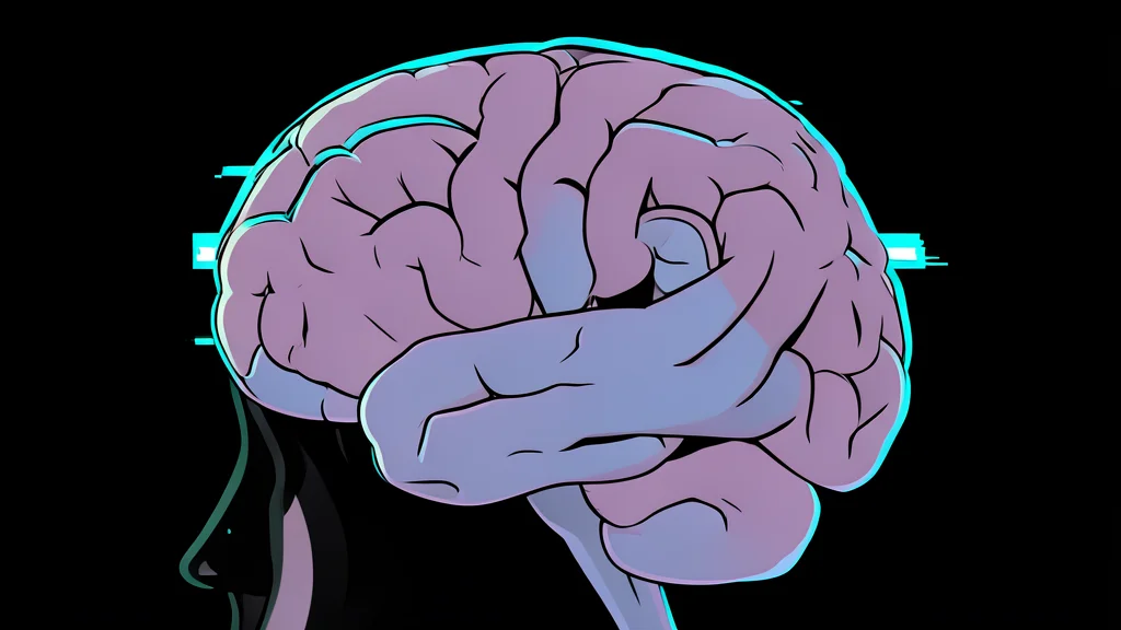
Researchers have identified a potential biological driver behind the persistent fatigue and cognitive issues that plague long COVID patients. A new study using advanced brain imaging suggests that the condition may cause physical damage to the brain's dopamine system, a network critical for motivation and movement. This finding offers a concrete target for future treatments in a condition that currently lacks evidence-based medical interventions.
Long COVID affects approximately five percent of the global population, with millions in Canada reporting symptoms that last months after initial infection. While these symptoms are well-documented, the underlying mechanisms in the brain have remained unclear until now. The new research, reported by ScienceDaily, connects specific brain injuries to the most debilitating symptoms experienced by survivors.
Imaging reveals distinct dopamine deficits
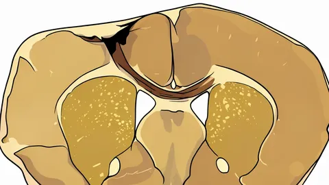
Scientists at the Centre for Addiction and Mental Health used positron emission tomography, a technique that measures biological activity in the brain, to compare patients with long COVID against healthy controls. They observed substantially lower levels of a specific marker for dopamine neuron health in the striatum, a region responsible for regulating motivation, movement, and thought processes. This indicates a reduced density of dopamine nerve terminals in these key areas.
The study found that the location of this damage correlates directly with specific symptoms. Lower marker levels in the ventral striatum were linked to a significant loss of motivation, while reductions in the dorsal putamen associated with slower movement. Memory difficulties appeared connected to lower levels in the caudate putamen. This pattern suggests that the physical injury to the dopamine system is not uniform but rather maps onto the specific functional struggles reported by patients.
Inflammation likely drives neuronal injury
These findings build on earlier work by the same team, which documented unusually high levels of inflammation in the brains of long COVID patients. That prior research showed inflammation was particularly pronounced in regions rich in dopamine-releasing neurons. The current study provides direct evidence that this inflammation may be causing permanent injury to the neurons themselves, rather than just temporary swelling.
By linking persistent inflammation to the observed loss of dopamine markers, researchers propose a causal chain: the virus triggers an immune response, inflammation follows, and that sustained inflammatory activity ultimately damages the nerve terminals. This mechanism explains why symptoms persist long after the acute infection has cleared, as the structural damage to the brain's reward and movement systems remains.
Targeting dopamine offers treatment hope
The identification of a specific biological deficit shifts the focus of long COVID research from general symptom management to targeted intervention. Since the damage is localized to the dopamine system, future therapies could potentially focus on protecting these neurons or compensating for their loss. This represents a significant departure from previous approaches, which often lacked a clear biological rationale for treatment strategies.
However, the trade-off is that this damage may be irreversible. If the neurons are permanently lost, treatments may need to focus on neuroplasticity or pharmacological support rather than restoration. Despite this challenge, the clarity provided by this imaging data allows for more precise clinical trials and a better understanding of why some patients struggle more with motor and motivational symptoms than others.