Human Brain Tissue Grows in Mice Lacking Cortex
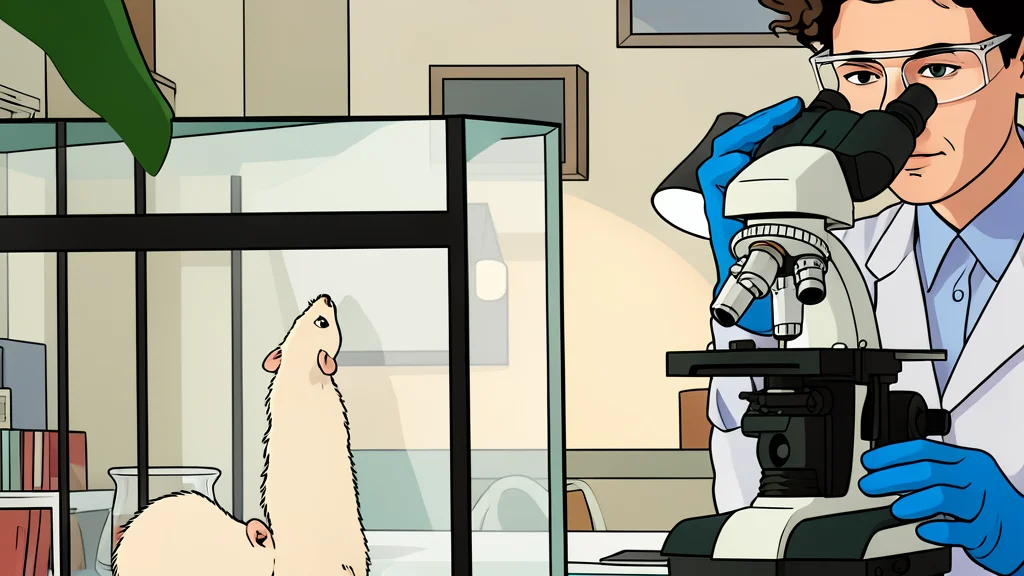
Stanford researchers have grown human brain cells inside mice that were engineered to lack most of their outer brain layer. The human tissue filled the space and connected with the host's nervous system, offering a new way to study human neurodevelopment.
Stanford researchers have successfully transplanted lab-grown human brain tissue into mice that were genetically engineered to lack most of their cerebral cortex. Within months, the human cells did not merely survive; they expanded to occupy more than 90 percent of the cortical space and formed functional connections with the mouse’s spinal cord and remaining brain regions. This breakthrough, reported by GN technics/space (en-US), provides a rare opportunity to observe how human neurons mature and respond to injury within a living, behaving animal.
The cerebral cortex is the outer layer of the brain responsible for complex thought, language, and decision-making. Studying this region has long been difficult because living human brain tissue is rarely available for research. While animal models have been useful, many biological features of the human cortex do not translate neatly from mice or rats, creating a significant gap in our understanding of human neurodevelopment and related disorders.
Engineered mice create space for growth
To overcome the limitation of competing native tissue, the team created a specific strain of mice known as apallial mice. These animals are genetically modified so that the starter cells for most of the neocortex never form. As a result, these mice have only about 2 percent of the cortical tissue found in ordinary mice, leaving a large cavity in the brain that can be filled by transplanted human tissue.
The researchers grew cortical organoids from healthy human donors and surgically placed them into the brains of two-day-old mouse pups. Over the next three months, the human tissue thrived and expanded. By the three-month mark, the majority of the cortical volume in these mice was human in origin, demonstrating that the tissue could integrate effectively without being outcompeted by native mouse cells.
Human cells integrate with host nervous system
The human neurons did not remain isolated in the cavity. They extended projections, formed synapses, and integrated with the mouse’s remaining brain regions and spinal cord. Behavioral testing showed that these mice performed broadly similarly to normal mice of the same age, though with subtle differences in gait and memory that hint at the stabilizing role of a full cortex.
This model offers a powerful tool for studying disease and injury. Because the organoids carry the donor’s genetic material, researchers can create mice whose human cortical tissue reflects the biology of a particular patient. This allows for personalized studies of neurodevelopmental disorders such as autism, epilepsy, and schizophrenia, providing insights that are difficult to obtain from traditional animal models or cell cultures alone.